What is sEMG?
The human body is a complicated network of neural pathways. For any movement, and for us to be able to interact with the world around us, the messages from the brain need to be successfully delivered to the muscles. Detecting and deciphering how the brain controls movement has continued to develop over the past years. A common way to assess human movement, and the contribution of different muscles towards force generation, is to use surface electromyography (sEMG or EMG). EMG is an experimental technique that involves the recording and analysis of the myoelectric signals formed by the physiological variations in the state of the muscle fibre membranes. By gaining insight into the status of muscular excitation, researchers, clinicians, sports practitioners, and engineers have been able to determine:
- The level of muscular effort involved – gained from assessing the overall EMG signal amplitude.
- Co-ordination between muscles – assessed by the activation timings of muscles.
- The effects of muscle fatigue – an exploration of the frequency component of the EMG signal.
- Neural control strategies – how is the brain adapting to training or external stimuli.
However, over the past decade there has been a rise in the popularity of using high-density surface electromyography (HDsEMG). In many applications, muscle activation may be treated as relatively homogenous across the whole region of the muscle. However, as we continue to evolve methodologies and technologies within electrophysiology, more evidence has come to light that muscles are heterogenous in their activation strategies between different regions of the muscle. This blog will look to provide further information on the background, applications, and current uses of HDsEMG within an array of scenarios.
The origins and outputs of HDsEMG
HDsEMG has been defined as the use of four or more closely spaced electrodes to give insights into the spatial distribution of myoelectric excitation across a muscle. While traditional EMG gives an insight into the global activation strategies of the muscles, HDsEMG offers a window into the regional excitation and hence the recruitment and modulation of localized motor units within or between muscles.
HDsEMG techniques have allowed for new questions to be asked within neurophysiology, neurorehabilitation, engineering, sports science, ergonomics, and clinical intervention assessments. Although the use of HDsEMG has grown over the past years, applications have been largely limited to constrained or isometric contractions. Now, with advances in fully wireless HDsEMG technology, applications of HDsEMG can include dynamic movements to provide unique insights from the neuromuscular system.
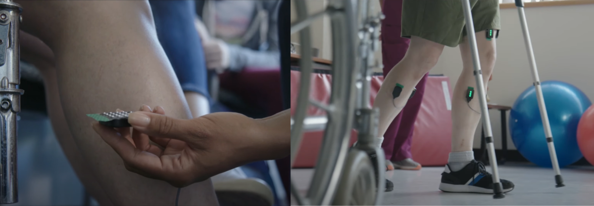

The non-homogenous patterns of excitation observed within muscles are reflected by distinct regions of the muscle behaving differently, and the reasons are multifactorial. Variations can depend upon the position of the joint, the duration and level of activation, or indeed the speed of contraction. It is therefore, in some circumstances, helpful to have a spatial representation of the EMG signal and an understanding f how this varies over time.
When assessing the signal at the surface of the skin, the amplitude is representative of the localized active muscle fibers in that area. Therefore, if muscle fibers, via the neural drive from the motorneurons, are preferentially recruited in certain areas of the muscle, the amplitude of the EMG signal at the surface of the skin will be characterized by this when shown on a topographical plot.
When employing a 2D HDsEMG grid electrode, amplitude variations can be accounted for in both the parallel and perpendicular direction of the motor unit action potential (MUAP) propagation. By recording the amplitude variations within the 2D area, we can assess how and why modulations of localized motor unit recruitment may occur to provide novel feedback on motor control and human movement.
Common Applications of HDsEMG
Whilst there are a wide variety of applications for HDsEMG within the assessment of the neuromuscular system, the following areas are some of the most common within the research field.
Pain and Fatigue
In healthy, or pain free humans, when undertaking a series of repeated or potentially fatigue inducing tasks, the muscle will redistribute activity to different regions of the muscle, hence minimizing the load on one particular region. This is a mechanism that is proposed to help reduce localized fatigue, or the onset of pain, as it was seen that patients with chronic musculoskeletal pain didn’t redistribute activity to different regions of the muscle throughout the duration of tasks.
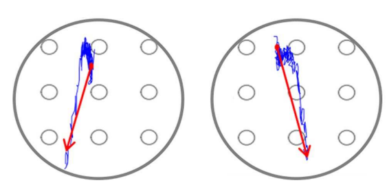
Centroid of a healthy control subject
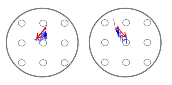
Centroid of a lower back pain subject

Graphics reproduced from Abboud, J., Nougarou, F., Pagé, I. et al. Trunk motor variability in patients with non-specific chronic low back pain. Eur J Appl Physiol 114, 2645–2654 (2014). https://doi.org/10.1007/s00421-014-2985-8
Patients and athletes who can mitigate against pain and/or fatigue tend to redistribute muscle fiber recruitment to different regions of the muscle during repetitive or sustained tasks. This enables them to still produce the sufficient force to complete the task, but without inducing localized overload on a specific region of the muscle.
By using HDsEMG, it is possible to assess if changes in regional activation occur which could minimize the effect of localized, persistent, and reoccurring pain or fatigue.
Sports Performance
Using HDsEMG within the application of sports performance can allow coaches, trainers, and practitioners to gain further insights into regional differences of muscle excitation that can further inform more targeted approaches to strength and conditioning programs or injury avoidance techniques.
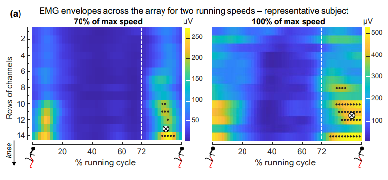
Graphic reproduced from Cerone GL, Nicola R, Caruso M, Rossanigo R, Cereatti A, Vieira TM. Running speed changes the distribution of excitation within the biceps femoris muscle in 80m sprints. Scand J Med Sci Sports. 2023;00:1-12. doi:10.1111/sms.14341
Preferential recruitment of differing regions within a muscle has been suggested as an adaptability of the neuromuscular system to avoid potential injury, from localized overload, or to increase neuromuscular performance. As an example, muscle-specific regional variations have been seen within hamstring muscles across the proximal and distal portions to aid performance and mitigate against injury inducing mechanisms during differing exercises.
Further studies looking at the effect of running speeds on regional activation (Vieira et al. 2023) found that, at speeds approaching 100% of max speed, the bicep femoris shifted activation more proximally during the late swing phase. It was suggested that, as the bicep femoris muscle approached full extension, activation shifted to help reduce potential for task failure or injury whilst also maintaining the necessary power output for sprinting.
Neurorehabilitation
Within different disease populations, changes in regional distribution have been seen to signify maladaptation from the associated mechanical or neurological deficit.
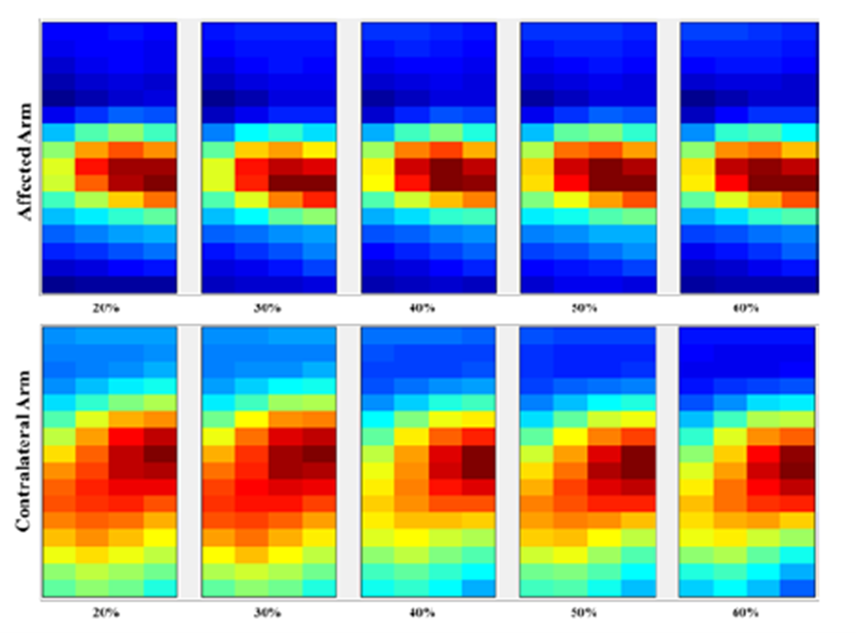
Graphic reproduced from Rasool G, Afsharipour B, Suresh NL, Xiaogang Hu, Rymer WZ. Spatial analysis of muscular activations in stroke survivors. Annu Int Conf IEEE Eng Med Biol Soc. 2015;2015:6058-61. doi: 10.1109/EMBC.2015.7319773. PMID: 26737673.
For instance, in stroke populations, studies have shown alterations in muscle architecture including the loss of muscle fibers and overall motor unit number reduction in paretic muscles of stroke survivors. Rasool et al. (2015) (Spatial analysis of muscular activations in stroke survivors – PubMed (nih.gov)) showed regional changes in amplitude clustering when comparing affected and unaffected limbs following a stroke.
The overall shrinkage of the area of activation that was seen in the affected side could likely be a sign of muscle atrophy. While the spatial pattern of the sEMG may refer to the mechanisms of motor unit recruitment within a muscle, the consistency of such patterns across force levels confirms the presence of mechanisms that do not change with the force requirements and hence the potential loss of motor unit numbers from de-innervation or disruption of the descending central nervous system pathways.
By using HDsEMG, disease progression or rehabilitation outcomes can be measured by assessing the regional distribution of the EMG signal and how this changes in relation to specific interventions.
Myoelectric Control
Multi-channel EMG sensors have been employed to provide a greater amount of input data for pattern recognition algorithms to create a more natural and intuitive human-machine interface. By using HDsEMG to decode the human motor intent, discrete patterns of regional muscle activation can be mapped to control outputs for the replication of specific movements of multi-functional prostheses with higher degrees of freedom.
When using the myoelectric signal from amputees for controlling an external device, HDsEMG offers a further option to increase the reliability of obtaining a functional signal from the residual limb. As amputees often have complications at the remaining limb due to changes in muscle architecture or muscle atrophy, a greater degree of spatial information may help in obtaining an increased understating of muscle activation.
Similarly, to widen the potential real-world applications of EMG within myoelectric control, there needs to be less expert knowledge needed to be able to work with the technology. Using standard bi-polar EMG to decode movement patterns for mapping within myoelectric control requires the electrodes to be placed on specific muscles. This may limit more novice users from being able to obtain accurate signals. Multi-channel EMG recording set-ups increase the spatial sampling of the myoelectric signal and hence put less emphasis on the placement of electrodes on particular muscles.
HDsEMG Research Example
HDsEMG in the past has been used to assess regional differences in muscle activation patterns during a variety of sporting activities, but little is known about the effects of altering the length of the muscular-tendon unit on the spatial distribution of motor unit recruitment during dynamic sporting activities. Current research, using 4 Trigno Maize HDsEMG sensors synchronized with the Xsens motion capture system, has investigated how altering the seat height during cycling effects regional activation strategies of the bicep femoris and medial gastrocnemius muscles.
Watch the following video to find out more and stay tuned for further findings.
