Key Takeaways
1
Myoelectric control is the broader term of using EMG signals as a control input. For prosthetics, these EMG signals are processed and classified into movements that are then translated into actuation of an external device.
2
Motor unit information from the amputated limb can be used to provide additional inputs to the controller algorithm. It was a common assumption that the neural activation between the residual limb and intact limb were the same when creating a model of muscle recruitment.
3
However, it was shown that alterations of MU properties exist within the residual limb which holds implications for prosthesis control as myoelectric models may need reparameterization to account for these neuromuscular changes.
What are myoelectric-controlled lower limb prostheses
Myoelectric control is the use of EMG signals to control actuation of an external device, typically a prosthetic. EMG signals are input into a processor and classifier that predicts the user’s intended movement, translating intent into an actuated motion. Accuracy of prosthetic actuation is largely dependent on the controller’s accuracy to decipher motor intent. The main goal of myoelectric control, in this instance, is to activate the prosthesis in a naturalistic fashion as an intact limb would.
Incorporation of Motor Unit Data
With the advancement of EMG sensors and EMG decomposition algorithms, researchers can now isolate individual motor unit (MU) information. MU information provides additional insights to the controller to create a better representation of intended movement. When creating a model of muscular recruitment to map onto an end-effector, there is a common assumption that neural activation of residual muscles and intact muscles are similar. However, recent research reveals that after amputations, the damage to the peripheral neuromuscular system disrupts afferent feedback and alters the internal representation of central motor control and EMG activity. Because of the disruption, assumptions about neural activation and corresponding EMG signals in residual muscles are questioned.
Current Research
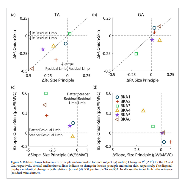
To get a deeper understanding of the altered neural and EMG activation states of below- knee amputees, the MU pool organization relations between intact and residual muscles was characterized by Noah Rubin and his team. MUs were determined through decomposing the EMG signals to determine the MU pool organization in a muscle. The MU was characterized by size principle, onion -skin, and rate-size association. The size principle refers to the size of the motor units, the onion-skin scheme refers to the concept that motor units activated at lower forces have greater firing rates, and the rate-size association refers to the firing rates of the motor units.
How EMG was used
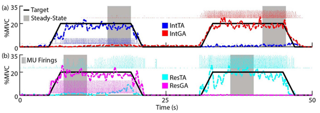
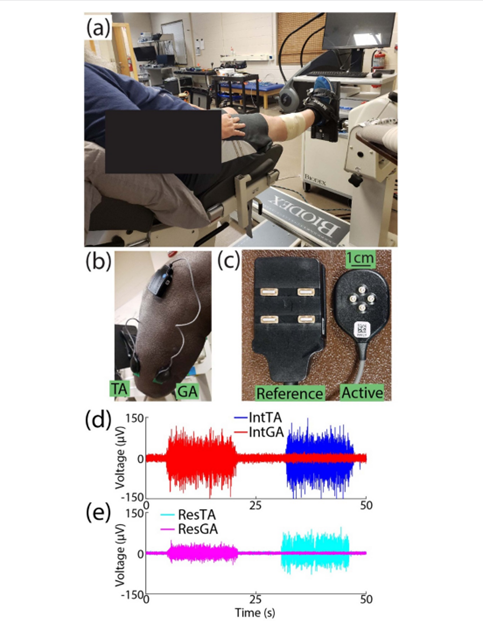
EMG was recorded on the tibialis anterior (TA) and the lateral gastrocnemius (GA) of both residual and intact limbs. sEMG signals were able to be acquired and decomposed through the Delsys Galileo sensor: four-pin array electrodes. A concern for the researchers while recording EMG signals was the possibility of crosstalk between the TA and the GA while subjects were alternating activation. However, this was not an issue for the researchers as the 5mm interelectrode distance of the four-pin array minimized the crosstalk between the muscles. The raw EMG signal was decomposed by the Delsys NeuroMap software to obtain the MU action potentials and timing of MU firings.
What was found
It was found from this study that disruption of the peripheral neuromuscular system, after limb amputation, changes the central neuromuscular control. In intact muscles, the size of the motor units’ amplitudes usually increase as the recruitment timeline increases. However, the residual TA exhibited less correlation between size and recruitment timeline. Regarding the onion-skin scheme, intact limbs typically display a negative correlation between motor unit mean firing rates and recruitment threshold. In the residual TA, that negative correlation is flattened. The rate-size association was unaffected in both residual limbs, but flatter decay rates were noticed. All these changes in characterization show that the neuromuscular control is much slower in the residual TA than in the intact TA. In addition, the changes are not consistent between muscles. The GA exhibited similar behavior to intact muscles. Despite that, there is still an interference in recruitment and discharge patterns of the MU pool and therefore EMG activity.
Future Impact
The purpose of this study was to test the assumption that the properties of EMG generation between residual and intact muscles were similar. EMG in this study was vital as the decomposed signals allowed MU information to be obtained and analyzed. The decomposed EMG signals gave insight into the activity of the neuromuscular system and convinced the researchers to reject the assumption. The evidence lay in the alteration of the size principle, onion-skin model, and rate-size association relationships. The difference in results between the two muscles showed that the neuromuscular control after amputation is not a consistent shift. In some muscles, there is a possibility for the relationships to stay the same. Regardless, if there is a shift in MU pool organization, it directly impacts the computational models of MU pool organizations and therefore alters simulations of EMG. For future research in EMG modeling, default parameterization of MU pool organization should not be assumed for residual muscles. The type of muscles and the alteration of correlative relationships between the size principle, onion-skin, and rate size association need to be taken into account for better representing motor intent for musculoskeletal model-based myoelectric controllers.
For more information on the use cases of EMG for lower-limb myoelectric control and human-machine interfacing, reach out to us at contact@delsys.com and learn more about the research area on Delsys Scholar.