From Lab to Life: Transforming Movement Studies with Dynamic TMS and EMG
Key Takeaways
1
During traditional TMS research, most studies take place with participants sitting still – far from the complex movements we make every day.
2
EMG recordings capture the muscles’ immediate responses to TMS pulses, offering a direct window into brain-muscle communication and providing an insight into action vs intention.
3
An innovative system at Cardiff University combines TMS and EMG during dynamic activities, revealing the intricate “conversation” between mind and muscle that powers our daily activities, bridging the gap.
Current Neurophysiology Research
The brain plays a central role in controlling the muscles in everything that we do, from the most dynamic and explosive sporting activities to the smallest of everyday tasks. Whilst biomechanical research into muscle activation in fundamental and sporting movements is incredibly common, neurophysiology research into how the brain connects to the muscles is less so. Given the central role the brain plays in controlling our muscles, research into understanding this connection is imperative for us to learn what, from a neural perspective, has led to a biomechanical change. To highlight some groundbreaking research in neurophysiology, we spoke to Jennifer Davies from Cardiff University to learn more about her innovative work into understanding how coordinated human movement is controlled.
We’re really good at describing changes that happen biomechanically, such as additional co-contraction around the knee if you’re in pain. But if we’re trying to target something like that from a rehabilitation perspective, we still don’t know from a neural perspective what has changed to cause this biomechanical change.
Dr. Jennifer Davies
Senior Lecturer within the School of Healthcare Sciences at Cardiff University
One tool that researchers like Jen can use to understand the connection between the brain and the muscles, is Transcranial Magnetic Stimulation (TMS). TMS is a non-invasive technique that uses magnetic fields to stimulate nerve cells in the brain, giving a picture of the connection between the brain and the muscles, the strength of that connection and which parts of the brain are responsible for moving certain body parts. TMS is an incredibly useful tool which provides a clean, direct stimulus during a movement, but without a way of detecting a readout of that signal at the muscular level, we’re unable to access the information from the stimulation and how a biomechanical change has been caused at a neural level. Jen uses Delsys EMG technology, in conjunction with TMS to receive a readout of that stimulation from muscle activation.
“EMG is critical because muscle activity is the readout of how responsive the neural system is. EMG is integral, TMS can’t tell us anything about the neural control of muscle without it”.
One of the issues with the application of TMS is that it is typically performed whilst sitting during isometric tasks, as the coil is heavy and needs to remain in a precise location to stimulate the desired part of the cortex. This means the findings don’t always translate to the world outside of the lab. However, Jen and her colleagues have created an innovative way to use TMS and EMG during walking, to allow us to further understand neural pathways involved in real-world movement.
Integrating TMS and EMG into a Dynamic Testing Environment
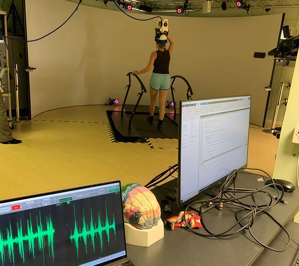
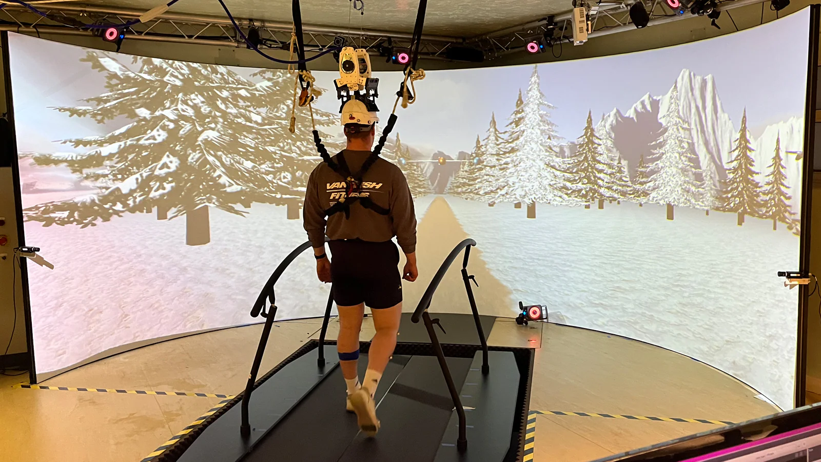
Participant completing an experimental protocol using the setup utilized by Jen and her colleagues.
Cardiff University, in collaboration with Magstim and Cardiff University Brain Research Imaging Centre (CUBRIC), have developed a novel and unique method of delivering brain stimulation during walking. The TMS coil is held in a helmet supported by springs, which holds the coil in a very specific position during movement. To ensure the coil is stimulating the exact location of the cortex required, the helmet has multiple points of adjustment to move up and down but also rotates to allow stimulation of the right or left hemisphere of the brain. As Jen’s research is completed during walking, the helmet needs to be able move side to side for participant safety but also to maintain the coil location, with an alert signalling if the TMS moves outside of 3mm from the desired location.
The TMS coil is integrated into a MOTEK Grail treadmill system, which allows for biomechanics of walking during different conditions such as incline/decline, and gait changes following perturbations to be tested. The Grail system also provides biofeedback during these tests to allow Jen to explore how the brain adapts to maintain balance and coordination with environmental and visual challenges, with a VICON motion capture system allowing for detailed analysis of the participants’ movements. Finally, Delsys Trigno Avanti sensors are attached to provide the critical real time EMG readout of how the nervous system responds to stimulation, and how the cortex alters the signal to deal with a wide number of scenarios.
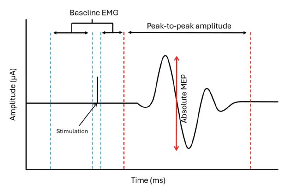
Figure 1. Graphical representation of simulation and muscle activity during data collection (from Huiberts et al. 2025).
Participants are introduced to the equipment and protocol to ensure comfort and confidence during the data collection. This includes a test stimulation to experience the feeling ahead of the protocol beginning and identify a threshold for muscle response using EMG. This test stimulation can be using TMS or peripheral nerve stimulation, depending on if the research is investigating cortex or spinal pathways respectively. Once setup and briefing are complete, the protocol to replicate the desired real-world scenario can begin on the treadmill. TMS is delivered at precise moments during the gait cycle where the brain and spinal cord play distinct roles in controlling muscle activation, such as heel strike, mid-stance and toe off. It is by delivering the stimulation at these moments, that allows Jen to assess how the nervous system responds dynamically, rather than in static conditions.
The setup and protocol have been carefully orchestrated and refined through its iterations to allow the strength and timing of neural signal to be measured. Trigno Avanti sensor are the final piece in the puzzle and by measuring peak-to-peak EMG amplitudes and normalising to pre-stimulation activity levels, accurate influence of TMS on the neuromuscular system can be monitored across different conditions. By comparing movement and EMG data across these conditions, Jen is able to uncover how neural pathways adapt to challenges.
“Delsys has been instrumental in letting us achieve the level of precision we need, especially when working in a dynamic and challenging environment like the treadmill.”
Implications of Dynamic TMS Outside of the Laboratory
“The really exciting thing is that this will allow us to test things that we haven’t been able to before, and the potential applications are really wide. In anything that the brain controls the muscles, which is essentially everything, this research can help us unpick that movement.”
As Jen and her colleagues have created such a novel and unique system to combine TMS and EMG, it has understandably taken a long time to develop and optimise. But after all that hard work, the value is clear, and research is now underway to help understand why dynamic biomechanical changes occur at a neural level. By enabling testing during dynamic activities like walking, the team has opened the door to studying how the brain and spinal cord coordinate muscle activity in real-world movements across a variety of conditions, such as fall prevention which is evidenced in the first study published using this system (Huiberts, Bruijn & Davies, 2025).
During this study, the team investigated how corticospinal excitability responded to mediolateral gait instability. They found that during lateral perturbation trials, participants showed increased step width and variability, with greater corticospinal excitability during unpredictable lateral destabilisations. Interestingly, the lower leg muscles did not see increased motor-evoked potentials (MEPs) whilst the upper leg muscles did, suggesting a greater reliance on these muscle groups for maintaining mediolateral stability. Corticospinal excitability was higher even during periods of low muscle activation, indicating that the brain is preparing for instability even before the muscles react. These findings provide key insights into how the brain adapts to balance challenges during walking, which can provide better understanding for rehabilitation strategies but also improve balance and prevent falls.
The dynamic environment ensures a high degree of real-world replicability that can be applied to multiple different areas across biomechanical, clinical and neurophysiological fields (Figure 2). Moreover, this research has the potential to transform rehabilitation strategies by providing deeper insights into neural pathways and their role in movement. These findings could inform targeted therapies that improve movement quality and reduce the risk of falls, particularly in populations with movement disorders or age-related challenges.
By combining TMS, EMG, and dynamic environments, Jen and her colleagues are transforming how we study movement and bringing laboratory science closer to real-world applications. As the system develops even further, for example by allowing the coil more freedom to move forwards and backwards in position on the treadmill, its applications will only become wider and stronger.
With so many areas of applications, the coming months and years will be a very exciting and busy time for Jen and the team as they complete research across these areas. Follow Jen’s progress to stay updated on her groundbreaking work and forthcoming publications, which are set to redefine how we study and treat neural and musculoskeletal conditions in dynamic, real-world scenarios.
For more information on the use of EMG as a quantitative assessment technique in neurophysiology, please reach out to contact@delsys.com.
References
Corticospinal Excitability in Response to Mediolateral Gait Instability
Huiberts, R., Bruijn, Sjoerd., Davies, J. (2025)
Explore More Topics
Client Spotlight
The Growing Importance of EMG and Human-in-the-Loop Strategies in Wearable Robotics