Inside Neuromechanics: Combining EMG and Imaging to Understand Human Movement
Key Takeaways
1
Using both EMG and ultrasound imaging gives researchers a more complete understanding of movement than either method alone.
2
Jeroen’s lab bridges the gap between fundamental research and real-world applications in sport performance, physiotherapy, and clinical care.
3
Research has shown that muscle activation patterns are unique to individuals, opening the door to more personalised rehabilitation and training approaches.
What is Neuromechanics?
In both sporting and clinical environments, understanding how our muscles generate force is fundamental, but many traditional biomechanical approaches are missing crucial pieces of the puzzle. While biomechanics has often focused on external motion and the forces produced by movement, these alone don’t fully explain how the body produces and controls force. To generate force, there must be muscle activation, but equally, there must be architectural changes within the muscle itself. Research will often measure muscle activation through electromyography (EMG) or muscle architecture through medical imaging individually, but at the intersection of these two areas, is the field of Neuromechanics. In relation to human movement, but in combination with EMG, we can get a more complete understanding and answer questions that we wouldn’t be able to without it. This is why Neuromechanics is such an important research area as it provides information on how our muscles activate and adapt from a mechanical perspective (fascicle length, muscle shape) and a neural perspective (activation, coordination patterns).
Often people are either studying the anatomy, the muscle side of things, or the neurophysiology. And I think we see more and more that by combining that, we can gain additional insights rather than just collecting data on both sides separately.
Prof. Jeroen Aeles
Assistant Professor of Biomechanics at VUB, Director of Brussels Neuromuscular Biomechanics Laboratory, Senior Academic Staff at Movement & Nutrition for Health & Performance (MOVE).
At the forefront of this field, is Professor Jeroen Aeles, Professor of Biomechanics at the Faculty of Physical Education & Physiotherapy at Vrije Universiteit Brussel (VUB). With an extensive background in muscle-tendon dynamics, neurophysiology and EMG, Jeroen and his colleagues are perfectly placed to combine muscle activation through Delsys EMG systems and mechanics, to unlock a deeper level of insight into movement control.
“You’ll often see people using ultrasound, using motion capture, but then often when you don’t have EMG, there’s so much missing information. Do we see an increase in muscle activation? A drop in neural drive? Are other muscles turning on and off, producing (counter) forces, affecting in turn the muscle you are studying?”
A Lab Built for Purpose
Jeroen established his own research team within the MOVE research group at VUB in 2023 with a clear vision: to bridge the gap between fundamental research and real-world application. Grounded in a neuromechanical approach, the lab enables Jeroen and his team to translate their understanding of muscle function and coordination into strategies that enhance performance, rehabilitation, and clinical practice.
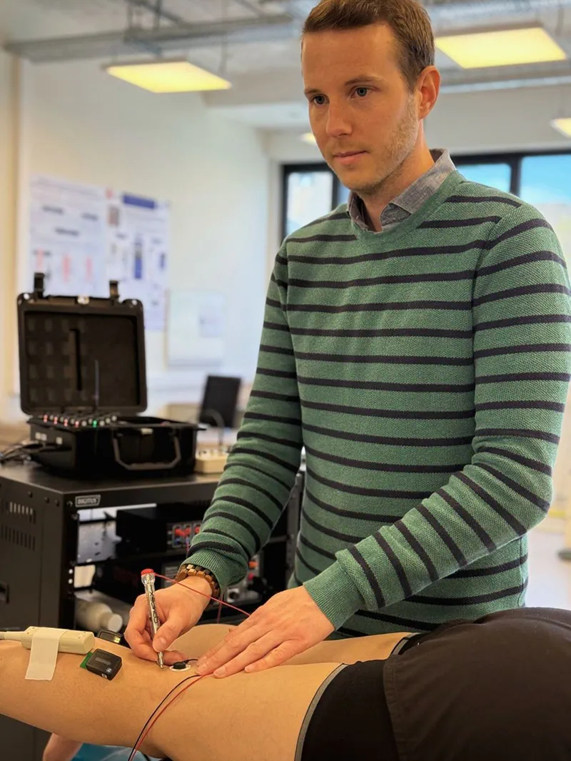
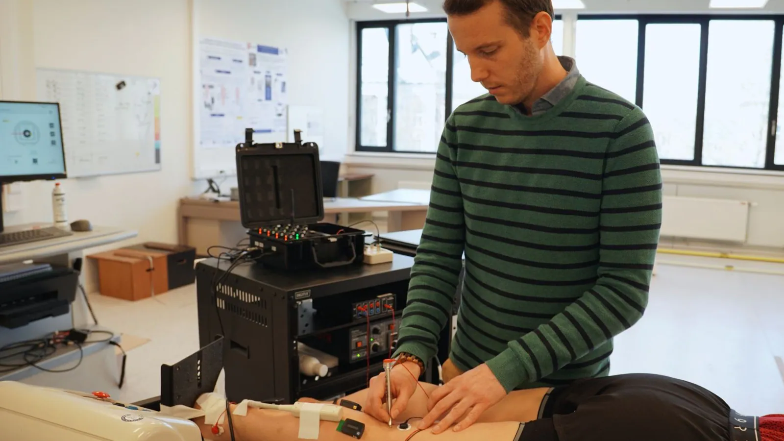
Jeroen completing experimental setup and identifying the optimal electrode placement for peripheral stimulation.
The lab’s research spans a diverse array of topics. These include reevaluating traditional EMG electrode placement guidelines, understanding how muscle morphology and function differ across individuals, and even species, and how these differences influence movement. At the core of every project is a rigorous, multimodal approach, seamlessly integrating tools like surface EMG, ultrasound imaging, and when needed, isokinetic dynamometry, motion capture, fine-wire or high-density EMG systems and peripheral or indwelling electrical stimulation. This flexible but consistent approach ensures that each study is tailored to the complexity of the question at hand, whilst maintaining scientific integrity.
“My go-to is classic bipolar EMG — it’s non-invasive and relatively easy to analyse. If I want to look at motor units, I’ll move to high-density EMG. If I need to look at a deeper muscle or a setup where space is an issue, I’ll use fine-wire EMG.”
And it’s not just the tools themselves that have been thoughtfully selected, but the physical design too. With studies spanning both the fundamental and applied domains, and involving health individuals, athletes and clinical populations the lab needed to be as adaptable as the research itself.
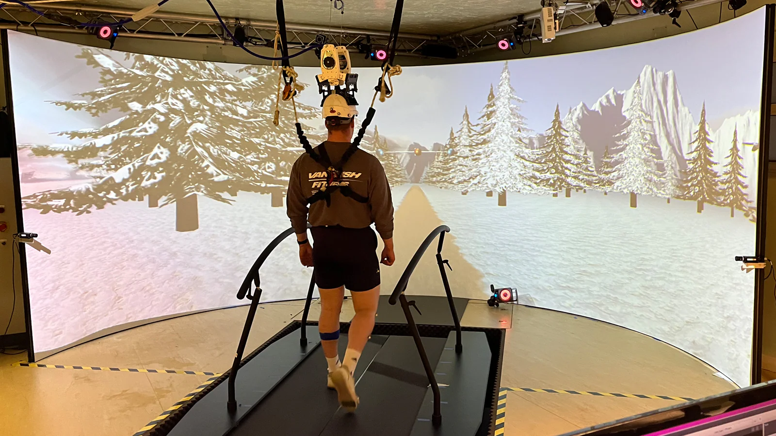
One half of the space is dedicated to a large, open area designed for dynamic movement tasks such as walking, running, and cycling. This is the heart of the lab’s sports biomechanics research. Here, the team utilise a Delsys Trigno Avanti system, alongside ultrasound imaging and motion capture technology, enabling real-time analysis of muscle activation and muscle architecture during movement.
“One of the reasons I bought the Delsys system is that I’d heard from other colleagues that it’s reliable and gives good data. And for the kind of work we do, where we’re often pushing the hardware to its limits, that really matters.”
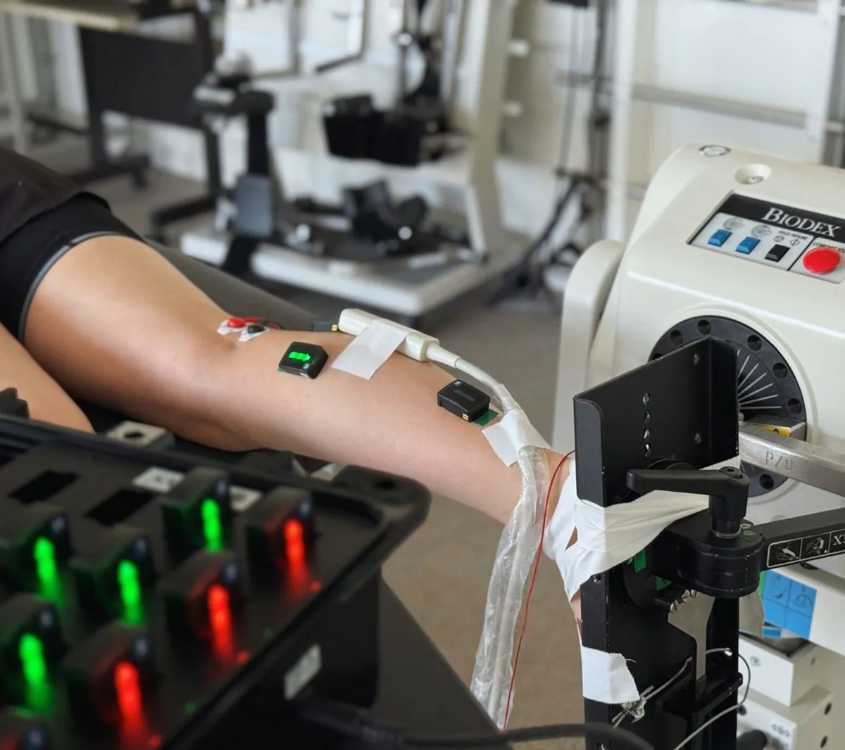
A typical controlled neuromechanics test in Jeroen's lab, combining EMG and ultrasound imaging of the triceps surae muscle group during testing with a Biodex dynamometer.
The second half is dedicated to controlled Neuromechanics testing, with a particular focus on the ankle joint, capturing the complexity governing triceps surae mechanics. Here, static and dynamic tests using a Biodex dynamometer are conducted to precisely manipulate and measure joint mechanics. As in the dynamic area of the lab, ultrasound imaging monitors muscle fascicle and tendon behaviour, while EMG captures muscle activation patterns. By combining these modalities, the team can investigate how neural and mechanical signals interact. This integrated approach allows Jeroen to determine whether observed changes in muscle activation are a cause or consequence of underlying mechanical shifts, providing deeper insight into the principles that govern human movement.
“In most experiments, the two most common things we use are EMG and ultrasound imaging. That’s our bread and butter.”
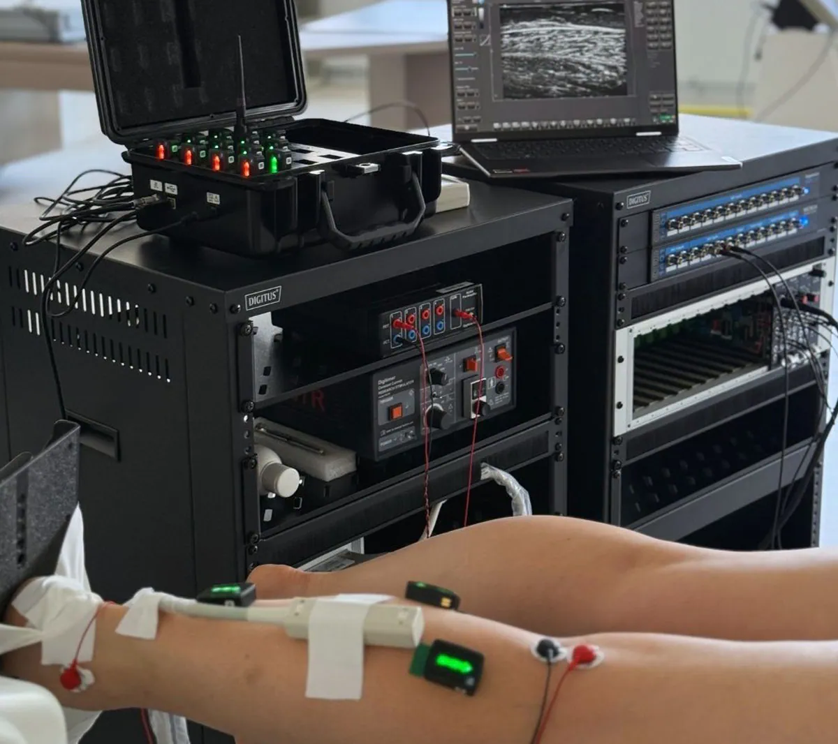
An experimental setup highlighting the hardware configuration and spatial constraints when using EMG and ultrasound together.
Research conducted in Jeroen’s lab often focuses on lower limb muscles such as gastrocnemius, soleus and tibialis anterior. The ultrasound probe will be placed as close as possible to the EMG sensor, whilst ensuring accurate spatial alignment and minimal signal interference. EMG sensor placement is critical to collect high quality signal, with minimised noise, movement artefacts and cross talk. The optimum location to achieve this is at the centre of the muscle belly. However, the ultrasound also requires a good image in the middle of the muscle, which means the probe is competing for space with the EMG sensor, especially in lower leg muscles. In some cases, there may be a blood vessel obstructing the ideal probe location, providing further complication. Whilst combining EMG and ultrasound imaging offers a wealth of complimentary information, the competition for space poses a big challenge.
To overcome this, using a wireless and compact EMG system will help greatly to not take up muscle surface area unnecessarily. More importantly, the team prioritises pilot testing and small validation studies to ensure signal quality, reliability and accuracy. In novel research such as Jeroen’s, these steps are essential to ensuring the data truly reflects what is happening inside the body.
“One of the things I really like about the Delsys system is that the sensor is integrated, so you don’t need to attach separate electrodes, amplifiers, or extra tape. It’s compact, wireless, and really easy to use, which is especially helpful when we’re competing for space with the ultrasound probe.”
Bringing Neuromechanics Into Sport & Rehabilitation
Jeroen and his team at VUB are conducting research that is both innovative and impactful. Their work is pushing the boundaries of how we understand individual muscle activation and coordination strategies, a fundamental step towards personalising approaches. Among their growing body of publications, one study really stands out as providing conclusive evidence that each person has a unique muscle activation signature during walking and pedalling (Aeles et al, 2021). This finding has far-reaching implications for individualised diagnostics, performance monitoring as well as bioinspired exoskeletons and the fundamental applications of electrical stimulation in certain muscular pathologies.
“With EMG data… we’ve actually shown that we can find these unique signatures in people’s muscle activation, which is pretty cool.”
Using Delsys surface EMG on eight lower limb muscles and applying machine learning with a novel interpretability technique called Layer-wise Relevance Propagation (LRP), the team showed that subtle features in EMG patterns such as amplitude, timing and coordination were consistent within individuals but varied between them. These activation signatures remained highly reliable across task conditions and test days which were at least 14 days apart, suggesting a strong reliability that these signals are physiological, rather than methodological artefacts. The machine learning algorithm identified where the unique identifier occurs, further evidencing the physiological correctness.
In most research, findings are presented as group averages to provide statistical power and ensure the results are applicable to the broader population. However, Jeroen and his colleagues’ findings suggest a need to shift our focus to individual data instead of relying solely on group averages, which is standard practice in clinics and sports, where training programs and nutrition is individualised. This opens the door to personalised rehabilitation, and performance monitoring, offering a deeper understanding of how individuals control movement. This work may also be applicable to clinical contexts by identifying signatures in people with injuries or neurological disorders.
“If we want to embrace this idea of personalised rehab or training, then research also needs to focus on the individual and EMG is central to doing that.”
To effectively utilise individual muscle activation profiles, robust protocols are essential for any future implementation in applied sport and clinical settings. As previously discussed, EMG sensor placement is critical. Traditionally, SENIAM guidelines have been widely used and referenced since the year 2000. With individual anatomy in mind, such commonly used guidelines must be accurate and up to date with current information. They must also be appropriate for clinical use and descriptive enough for practitioners and clinicians who are unfamiliar with anatomy to find the correct sensor location without ultrasound. Jeroen and the team have already begun to look at the current guidelines, answering the above questions and create new recommendations and guidelines which are in line with current information.
Multi-disciplinary research, such as Neuromechanics, is incredibly valuable for advancing our understanding of human movement. Jeroen’s published and upcoming research truly highlights his passion to advance this field and push the boundaries of how we measure, interpret, and apply muscle activation data. His work not only challenges long-standing EMG methodologies but also paves the way for more precise, individualised approaches to sports science and rehabilitation.
You can explore more of Jeroen’s publications on ResearchGate, where his publications highlight the depth and breadth of his team’s contributions to the field.
For more information on the use of EMG as a quantitative assessment technique in Neuromechanics, please reach out to contact@delsys.com.
References
Revealing the unique features of each individual's muscle activation signatures.