Research & Innovation based on the research by Dr. Pieter Bas de Witte
Background
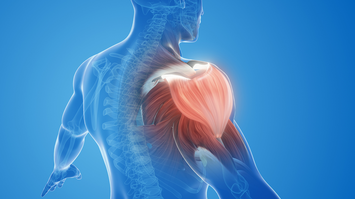
Subacromial Impingement Syndrome (SIS) and Rotator Cuff (RC) tendon tears are frequently diagnosed in patients with shoulder pain. Both conditions show similar symptoms, including pain with arm abduction, although symptoms are generally worse in patients with RC tears. Some report that both are stages of the same condition, where SIS may progress to a RC tear due to degeneration. Their etiology is unclear and often reported as heterogeneous. Therefore, clinical decision making is complicated, and treatment strategies and their results vary greatly. New methods are needed to investigate whether there are actually various underlying mechanisms for these diseases, that might need specific treatment approaches.
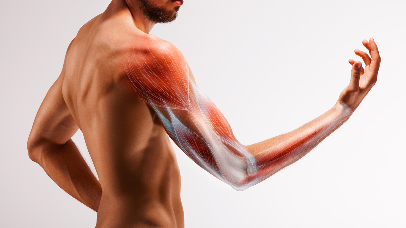
Electromyography (EMG) offers the opportunity to study muscle activation patters around the shoulder. However, the activity of RC muscles is hard to measure directly and generally requires nerve-blocks or intramuscular EMG electrodes. Therefore, methods to measure RC function indirectly, by assessing adductor co-activation and Deltoid activation, were developed at the Laboratory for Kinematics and Neuromechanics (LK&N) at LUMC. These studies have shown that RC tear patients have pathologic ADductor activation, during arm ABduction (pathologic “co-activation”), presumably in order to “pull” the humeral head down during abduction, in order prevent encroachment (i.e. impingement) of RC tendons. Also, a method was developed to express adductor co-activation in an easy-to-interpret measure: Activation Ratio (AR, -1<AR<1), where lower values mean more co-activation.
Possibly, adductor co-activation can play a role in unraveling the underlying mechanisms of SIS and RC tears, in order to improve diagnostics and treatment strategies.
keywords: muscle fatigue, electromyography, mean frequency, accelerometry

Pieter Bas de Witte, MD, BSc
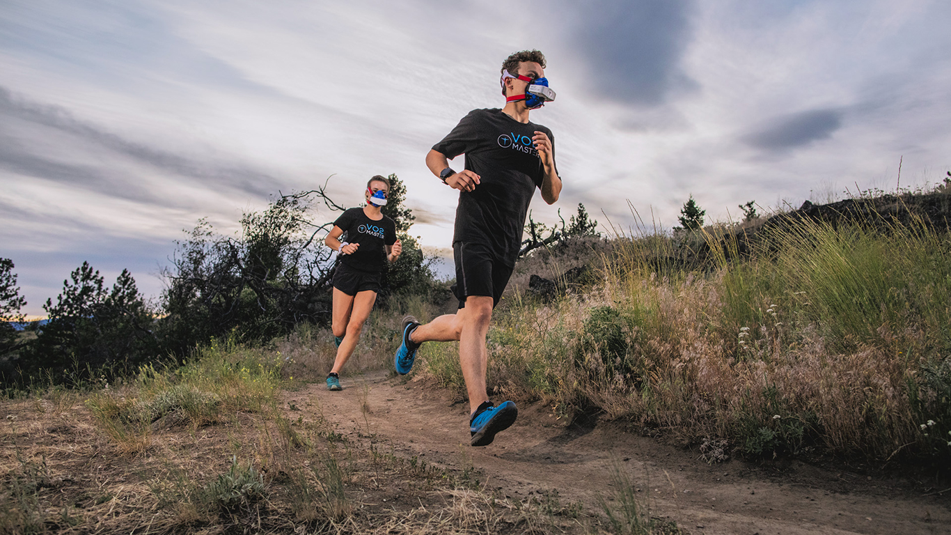
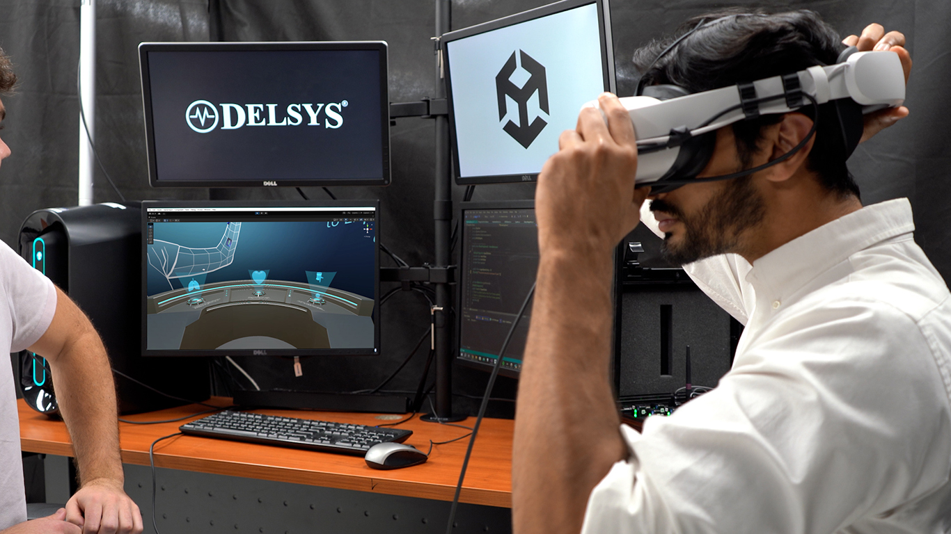
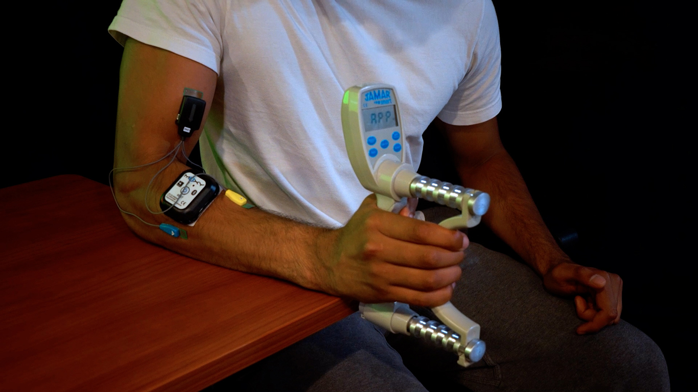
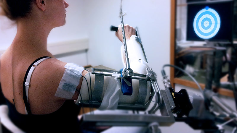
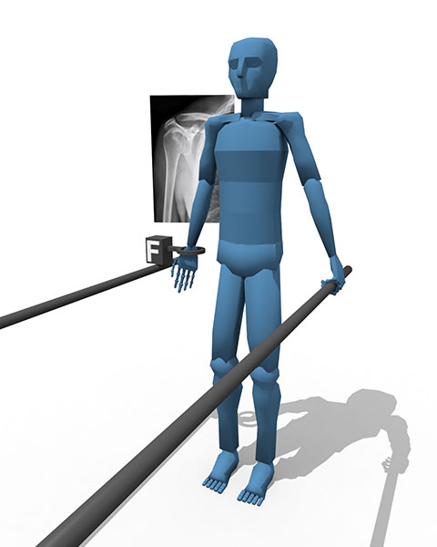
Since 2009, Dr. Pieter Bas de Witte has been active in several clinical and biomechanical studies at the department of Orthopaedics at LUMC. He has applied the EMG-methods that were designed at LK&N and developed them further for use in healthy subjects, patients with SIS and patients with RC tears. For this purpose, the original set-up (Figure 1) was developed into a more mobile set-up (Figures 2&3), in order to combine analyses of muscle activation patterns, with shoulder radiographs acquired during active tasks.
Research and Innovation
In a first study, investigating Supraspinatus function with intra-muscular electrodes in healthy subjects, we found that the function of the Supraspinatus is significantly more variable than the function of the Deltoid1.
In a second study, investigating adductor co-activation, a healthy control group had adductor AR’s of 0.7-0.9 with good test-retest reliability. RC tear patients showed significantly more adductor co-activation during abduction, with AR’s ranging from 0.3- 0.5, discriminating RC tear patients and healthy controls2.
In a third study, the same set-up was used, but now with simultaneously acquired shoulder radiographs and in RC tear patients, SIS patients and controls. The results showed that at rest, the space between humerus and acromion is significantly smaller in RC tear patients compared to SIS patients and controls. During abduction, cranial humerus translation was observed with equal magnitudes for patients (RC tear and SIS) and controls. EMG measures showed pathologic “out-of-phase” adductor co-activation during abduction in specifically the RC tear patients. However, no association between adductor co-activation and humerus cranial translation was found3.
In a fourth study, we found increased (compensatory) Deltoid activation in patients with RC tears. This Deltoid compensatory role was reduced one year after surgical RC cuff repair, suggesting (partially) restored RC function4.
Conclusion
The studies by Dr. de Witte show that biomechanical measures (EMG measure with Delsys equipment) can be applied in easy-to-use set-ups with interpretable outcome measures, in order to study underlying mechanisms for shoulder symptoms. This gives clinicians more insight in these mechanisms and might help in developing diagnostic methods and flowcharts in order to optimize clinical decision making and treatment outcome.
References
- de Witte PB, Werner S, Ter Braak LM, Veeger HE, Nelissen RG, and de Groot JH. The Supraspinatus and the Deltoid – Not just two arm elevators. Hum Mov Sci. 2014 Feb;33:273-283
- de Witte PB, van der Zwaal P, Visch W, Schut J, Nagels J, Nelissen RG, and de Groot JH. Arm Adductor with arm Abduction in rotator cuff tear patients vs. healthy – Design of a new measuring instrument. Hum Mov Sci. 2012;31:461-471.
- de Witte PB, Henseler JF, van Zwet EW, Nagels J, Nelissen RG, and de Groot JH. Cranial humerus translation, deltoid activation, adductor co-activation and rotator cuff disease – different patterns in rotator cuff tears, subacromial impingement and controls. Clin Biomech (Bristol, Avon). 2014 Jan;29(1):26-32.
- de Witte PB, van der Zwaal P, van Arkel ER, Nelissen RG, and de Groot JH. Pathologic Deltoid activation in rotator cuff tear patients: normalization after cuff repair? Med Biol Eng Comput. 2014 Mar;52(3):241-2499