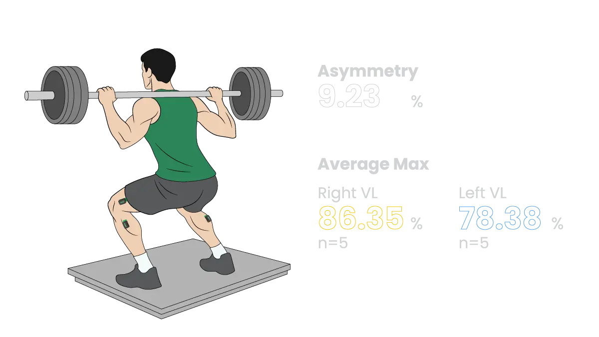
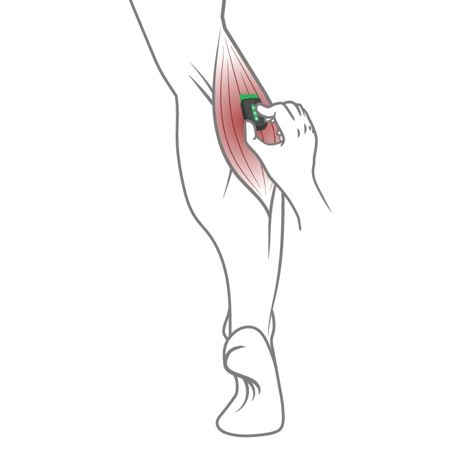
Placement must be consistent and anatomically precise. Guidelines like SENIAM help standardize this, but practitioner training is key.
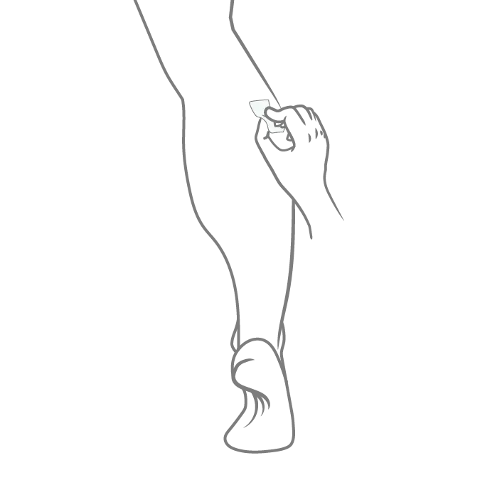
Shaving, cleaning, and prepping the skin reduces impedance and improves signal quality. Poor preparation can lead to noisy and unreliable data.
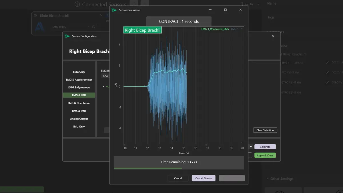
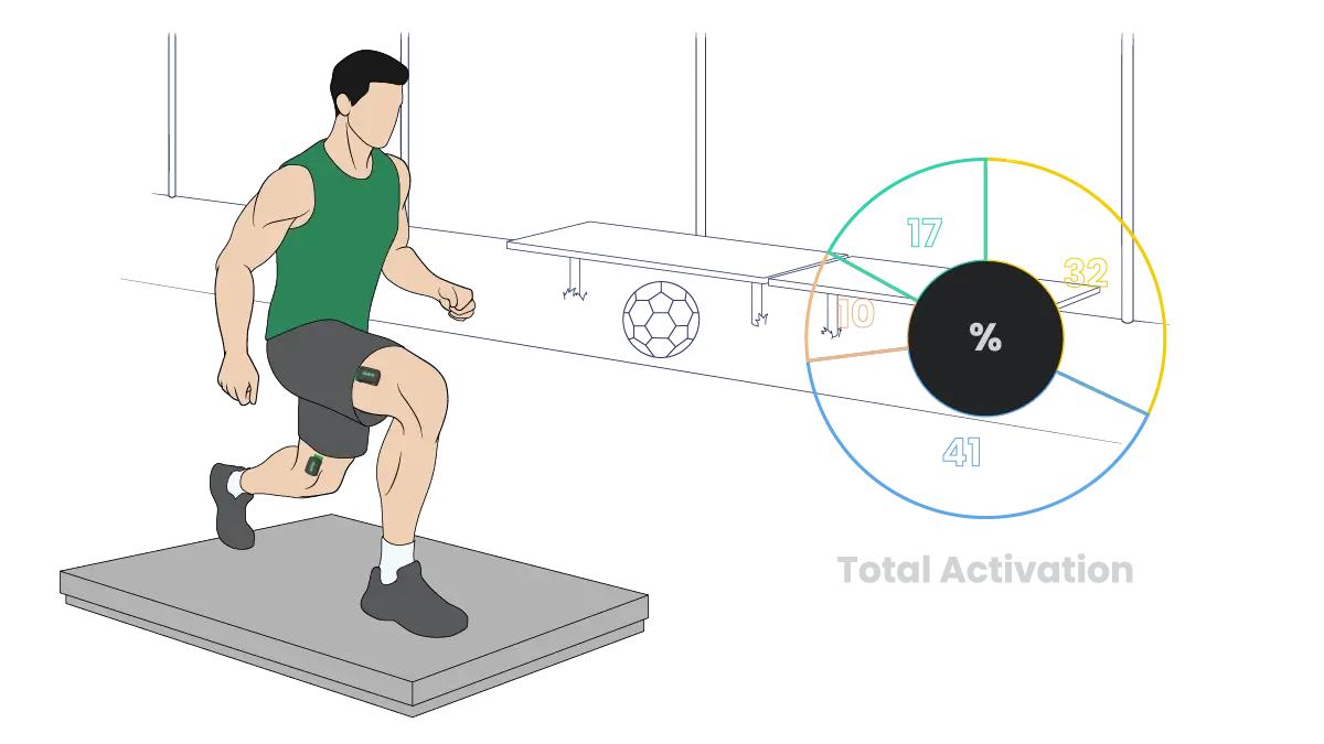
Comparing EMG amplitude across sessions or athletes requires a reference. This is often a maximal voluntary contraction, but standardized submaximal tasks can also work depending on context.
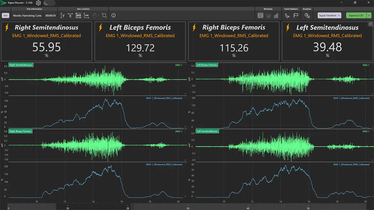
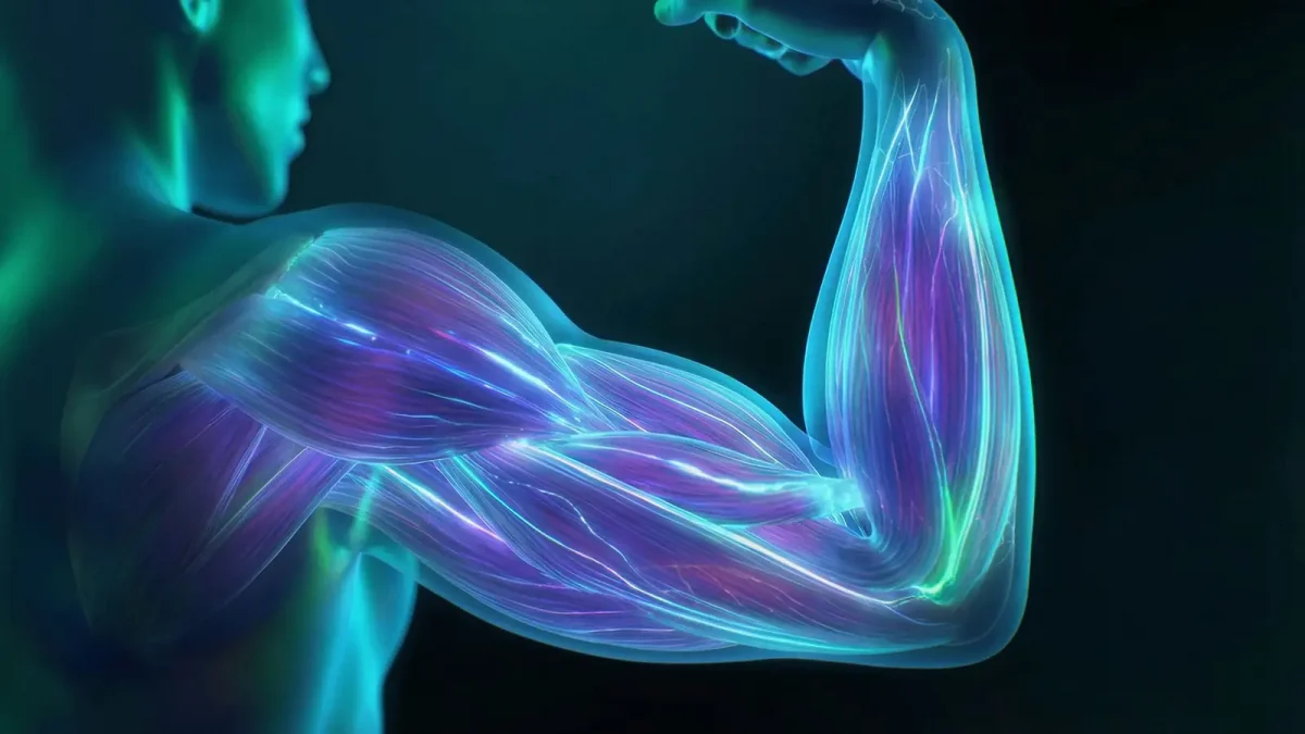
EMG does not equal muscle force. Signal amplitude reflects neural drive, which interacts with factors like muscle length, fatigue, and electrode position. Always interpret within context.
Shaving, cleaning, and prepping the skin reduces impedance and improves signal quality. Poor preparation can lead to noisy and unreliable data.
1 of 5 2 Electrode Placement
Placement must be consistent and anatomically precise. Guidelines like SENIAM help standardize this, but practitioner training is key.
2 of 5 3 Normalization Comparing EMG amplitude across sessions or athletes requires a reference. This is often a maximal voluntary contraction, but standardized submaximal tasks can also work depending on context.
EMG does not equal muscle force. Signal amplitude reflects neural drive, which interacts with factors like muscle length, fatigue, and electrode position. Always interpret within context.
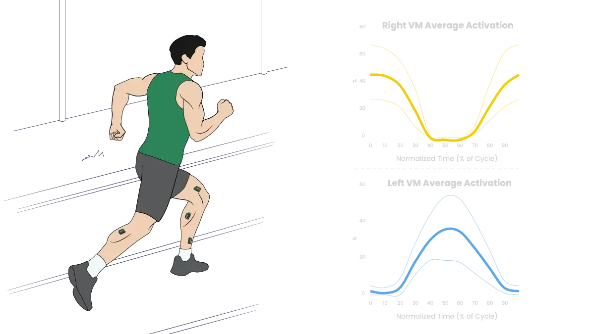
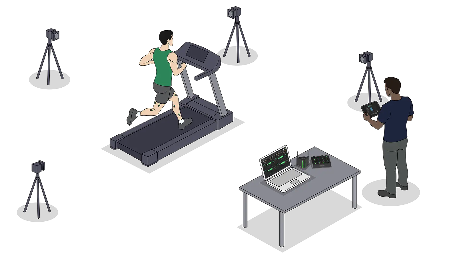
4 of 5 5 Data IntegrationEMG is most valuable when combined with kinematics (motion capture), kinetics (force plates), or other wearable sensor data. Alone, it gives partial insight; together, it builds a complete picture.
5 of 5